State regulation of neuromodulatory control in the neural network
Overview
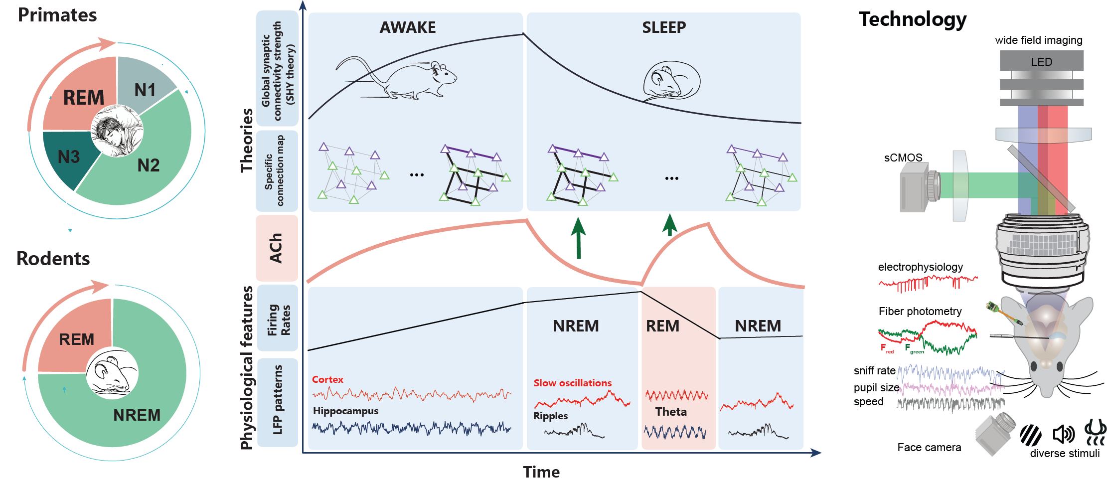
How does the sleeping brain decide which experiences are worth keeping? The synaptic homeostasis hypothesis (SHY) proposes that sleep restores homeostasis through global synaptic downscaling — yet sleep is not a uniform state. Across the NREM–REM cycle, acetylcholine (ACh) oscillates dramatically: suppressed during NREM slow oscillations and hippocampal ripples, then surging to an isolated peak during REM theta. We study how these ACh dynamics serve as a phased control signal that drives global downscaling in NREM while selectively reinforcing learning-relevant synaptic connections during REM — translating sleep architecture into the selective long-term storage of memory.
System Architecture
The platform is built around a learning–post-learning sleep paradigm that coordinates three recording modalities with circuit-level temporal resolution.
Fiber Photometry
Real-time readout of bulk neuromodulator signals (ACh, dopamine) and calcium activity from genetically defined cell populations during behavior.
Electrophysiology
Multi-site silicon probe recordings capturing single-unit and LFP activity across hippocampal and olfactory circuits simultaneously with behavior.
1P Imaging & Stimulation
One-photon calcium imaging and optogenetic stimulation with targeted single-cell resolution, phase-locked to the sniff cycle.
Behavioral Paradigm
Animals are free-moving and perform T-maze task:
| Event | Timing |
|---|---|
| T-maze Task | Alternating trials with left/right arm choices, using visual/auditory cues |
| Reward | Water delivery on correct lick |
| Laser trigger | State-locked — fires at REM onset/offset (\(t_{stim}\)) |
state detection is critical — it aligns neural activity to a physiologically meaningful reference and enables comparison across trials and animals.
Continuous Monitoring Streams
Beyond sleep-stage classification, we continuously record four parallel signals:
- ACh — cholinergic tone via fiber photometry, capturing the NREM–REM oscillation cycle in real time
- LFP — local field potentials from hippocampus and cortex, resolving slow oscillations, sharp-wave ripples, and theta rhythms across sleep stages
- Wide-field calcium imaging — mesoscale cortical activity via sCMOS, tracking spatiotemporal reactivation patterns across the wake–sleep transition
- Behavioral state — sniff rate, pupil size, and locomotion speed as continuous arousal and state-transition markers
This multivariate state vector allows us to precisely assign each neural event — a ripple, a reactivation, a firing rate change — to its position within the NREM–REM cycle and the corresponding cholinergic phase, turning correlative observations into a causally interpretable record of how ACh orchestrates memory consolidation during sleep.
All modalities are referenced to a common hardware clock. Sleep-stage transitions, optogenetic perturbations, and calcium or photometry frames are co-registered to the same timeline, enabling precise alignment of ACh dynamics, network oscillations, and behavioral state at the resolution of individual sleep cycles.
Interested in collaborating or accessing our behavioral platform? Get in touch.